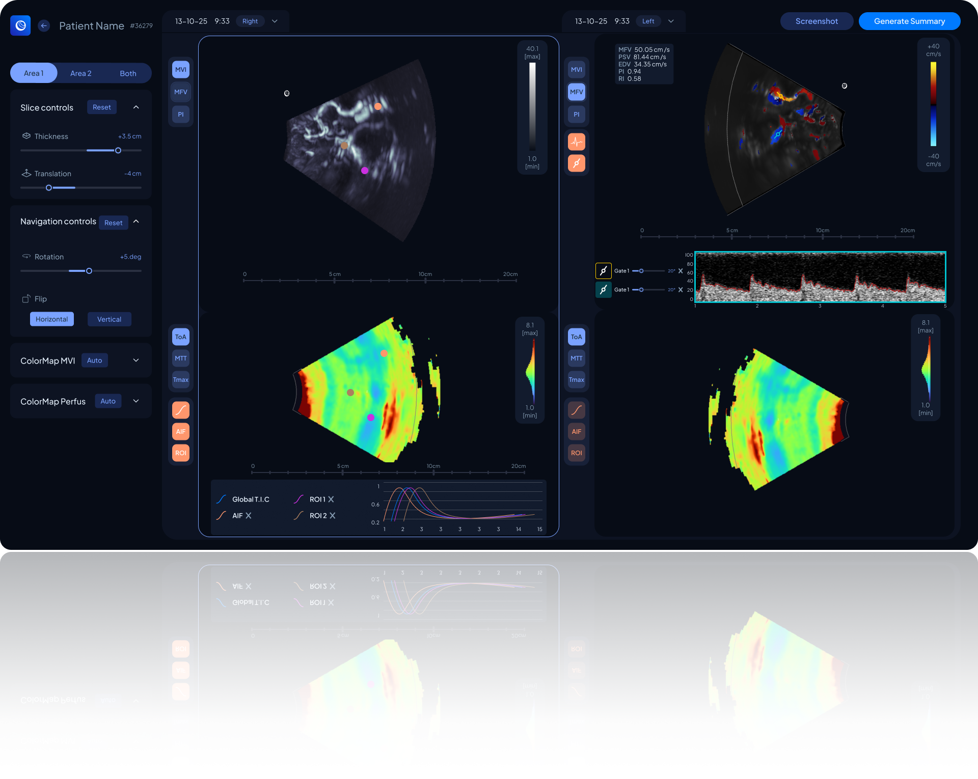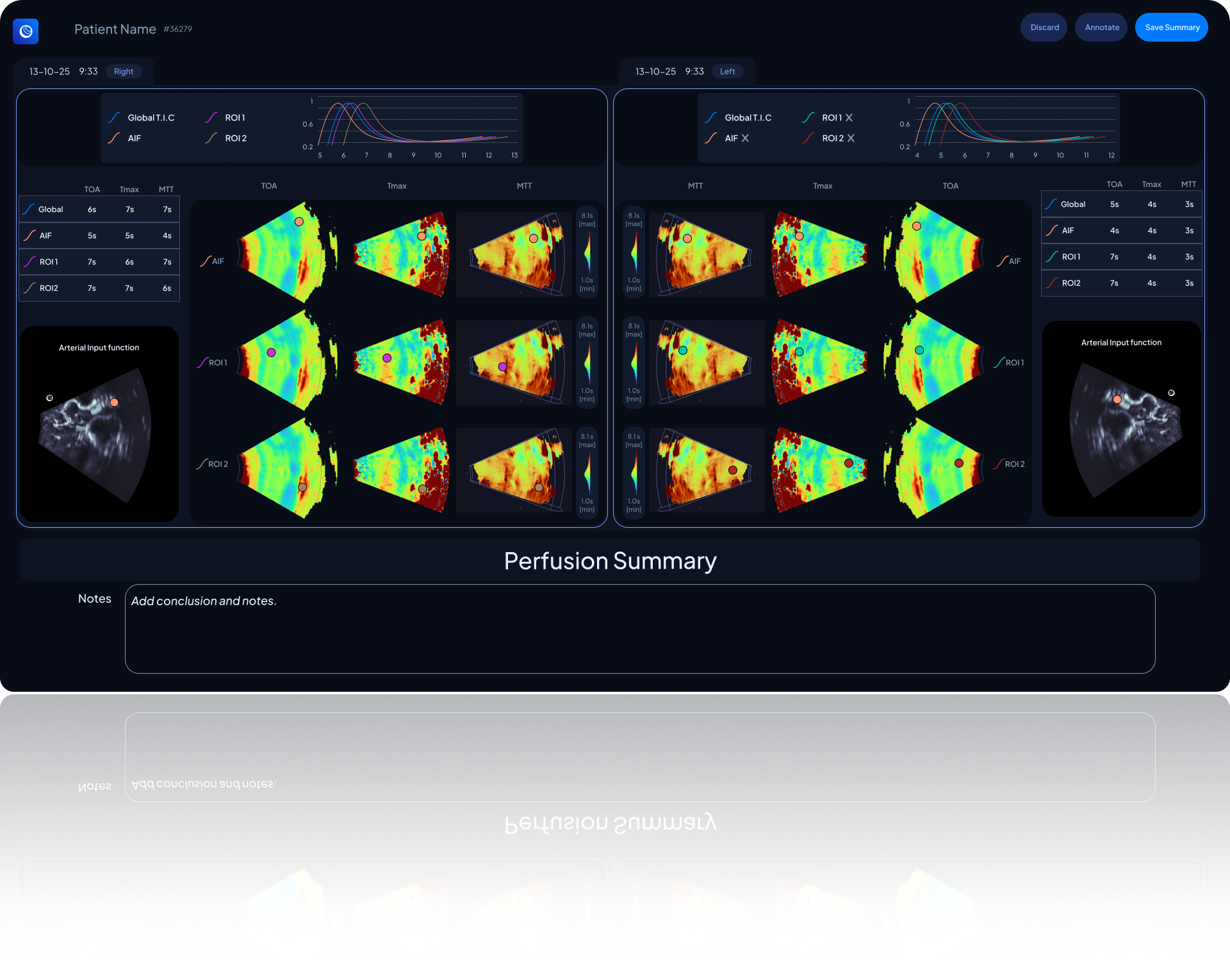
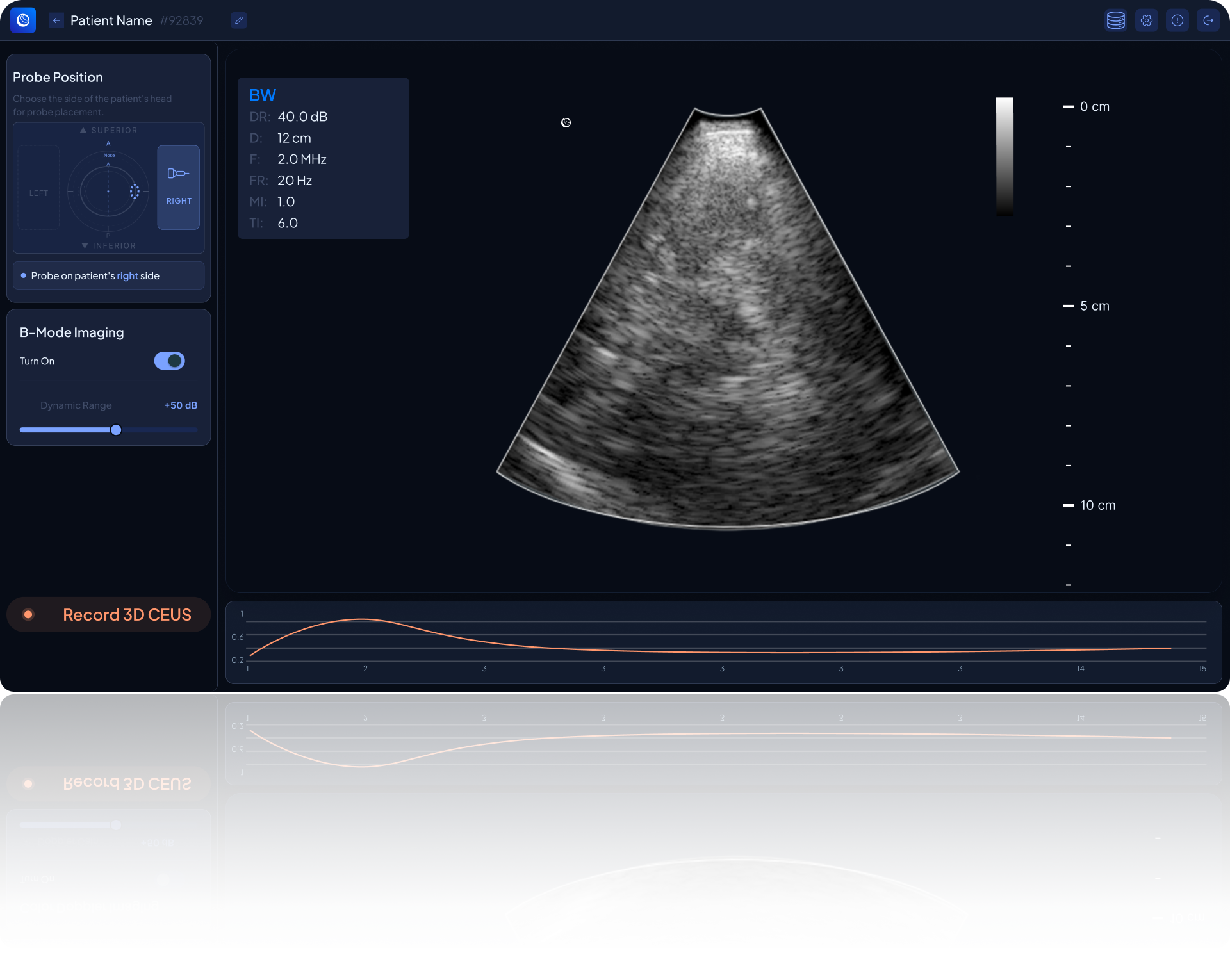
A conventional 2D grayscale ultrasound imaging mode delivering anatomical visualization.
Used as a baseline reference layer to ensure precise positioning before initiating Resolve Stroke’s proprietary 3D CEUS sequence.
Images for illustrative purposes only.
Bring imaging
closer to patients’ needs
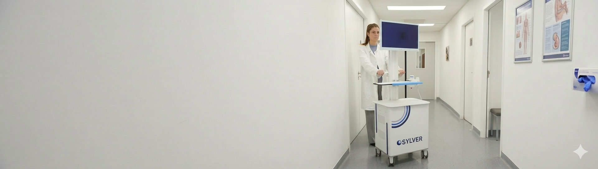
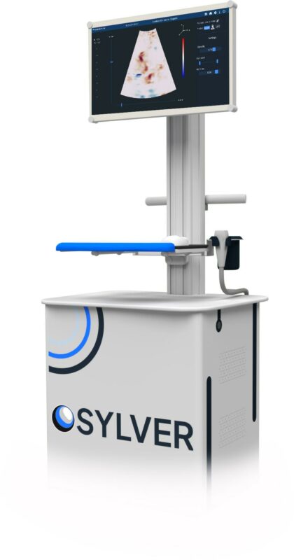
RS Views embedded on a first Ultrasound Cart