Ultrasound is the next frontier for AI in medical imaging
AI transformed MRI & CT
Structured databases, standardized formats, and massive labeled datasets made these modalities AI-ready. The result: faster and better diagnosis and workflows, and billion-dollar AI models. But MRI & CT reach less than 20% of patients globally.
Ultrasound, #1 imaging modality, underexploited
Today, conventional ultrasound remains limited due to performance constraints and user dependency, with only 10% of the data being utilized. Hardware breakthroughs have unlocked a new asset: ultrasound data at scale. The bottleneck is no longer the probe, it’s the software.
Empowering medical decisions with Ultrasound
OUR AMBITION
As ultrasound hardware commoditizes, the value shifts to data and algorithms.We build the AI software layers that turn this commodity hardware into a clinical intelligence platform, extracting biomarkers that were invisible until now.
OUR FIRST APPLICATION
Unlocking the brain with Ultrasound
The brain has been out of reach for ultrasound until now. We apply AI to transcranial signals to extract biomarkers in real time, directly at the bedside. First targets: neurology, intensive care, and emergency medicine.

Bring change to all stakeholders
Our clinical ambition*
Aiming for faster diagnosis and earlier treatment, helping reduce irreversible injuries and improve outcomes.
Our operational ambition
Supporting faster decisions, safer workflows, and better resource allocation.
Our economic ambition
Targeting cost‑neutral adoption, scalable long‑term savings, and alignment with value‑based care models.
*Clinical outcomes are under evaluation.
From imaging
to predictive insights
Creating change together


Our latest news
Resolve Stroke connects with global experts
Over the past few months, the Resolve Stroke team has been actively connecting with leading experts in neurology and neurosurgery across North America and Europe:

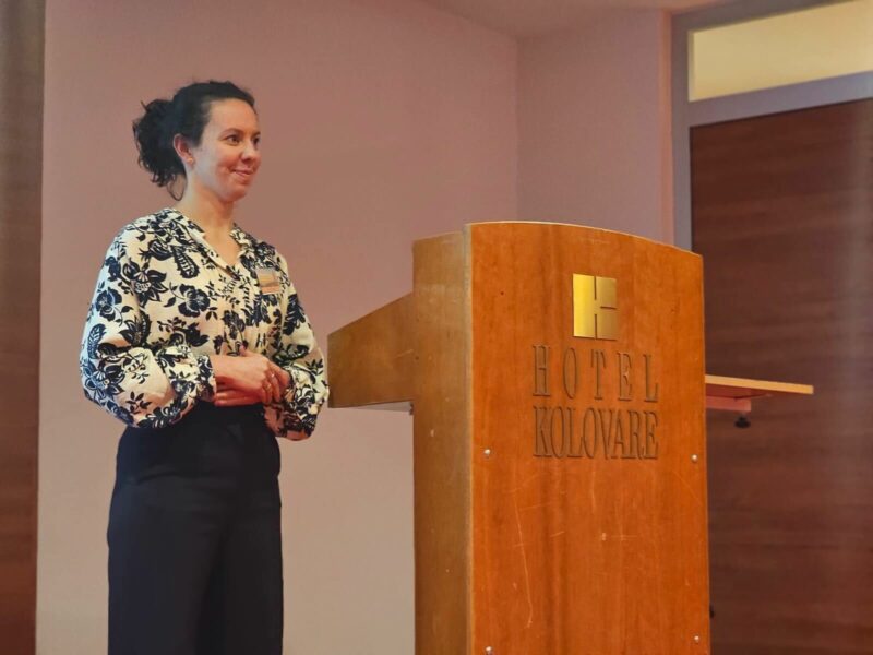
Prix Galien 2025 Official Selection
Resolve Stroke is honored to be officially selected for the 2025 Prix Galien France in the Medtech & Digital Solutions category. This prestigious nomination highlights the impact and potential of our ultrasound-based neuroimaging technology to advance patient care. We thank the Prix Galien jury for recognizing our commitment to innovation in healthcare.
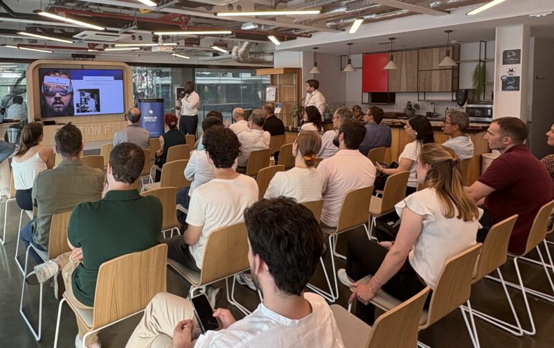
Resolve Stroke hosts neuro-ultrasound workshop at Station F
Resolve Stroke organized a hands-on workshop at Station F, bringing together clinicians and researchers interested in the future of ultrasound in neurocritical care. Participants discovered our SYLVER platform and RS Suite, joined expert-led discussions, and explored how software-driven ultrasound can unlock new possibilities for brain monitoring at the bedside. Thank you to all who joined!
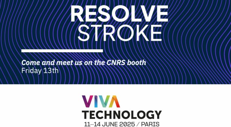
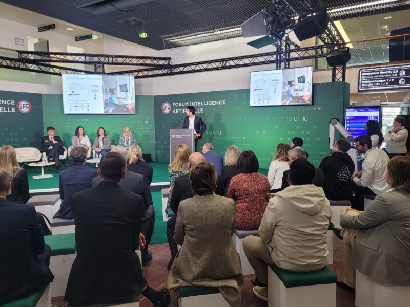
VivaTech 2025
Resolve Stroke was invited to pitch on stage at VivaTech 2025 in Paris, thanks to the support of CNRS. We were thrilled to present our innovations in health tech and to connect with industry leaders at this major international event.
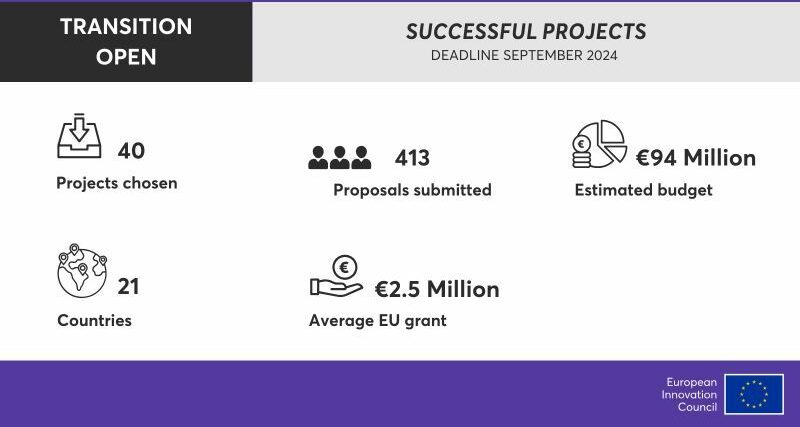
2.5M€ EIC Transition Grant Award
We are proud to announce that the RS-NeuroMarkers project has been selected by the European Innovation Council for a prestigious EIC Transition Grant. This €2.5M grant recognizes our mission to advance point-of-care neuroimaging, making brain monitoring more accessible through ultrasound technology. With RS-NeuroMarkers, we will integrate clinically validated brain biomarkers into our software engine, helping clinicians detect and monitor cerebral pathologies directly at the bedside.
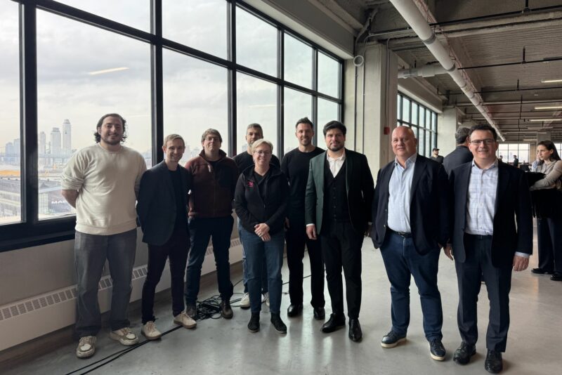
Resolve Stroke Pitch at NYC Deep Tech Week
Our CTO Vincent Hingot took the stage at Deep Tech Week in New York City to present Resolve Stroke’s vision for the future of neuroimaging. Vincent shared how we leverage ultrasound physics and advanced signal processing to deliver real-time brain insights. We’re honored to be part of this vibrant community of innovators shaping the future of medicine.
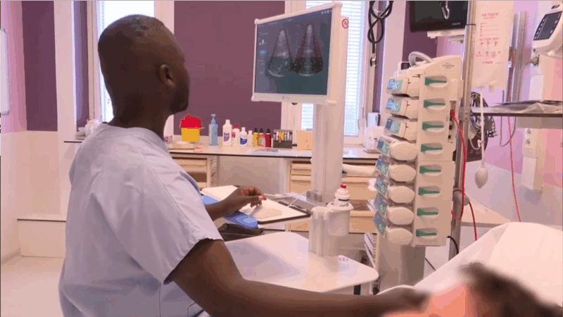
Bedside brain perfusion imaging: Dr. Gakuba showcases SYLVER platform
In a recent video interview published by Le Quotidien du Médecin, Dr. Clément Gakuba (CHU Caen) highlights how our SYLVER ultrasound device enables advanced brain perfusion imaging directly at the patient’s bedside. The technology was evaluated in a first clinical study involving 15 patients with subarachnoid hemorrhage, demonstrating both feasibility and the ability to identify different perfusion profiles.
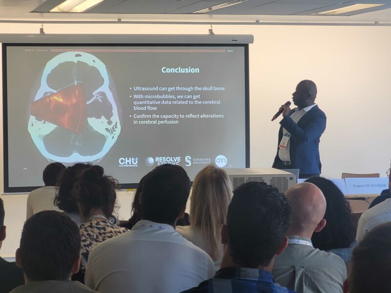
Clinical trial – First results
First results from the collaboration between CHU Caen Normandie and Resolve Stroke officially presented at the SFAR meeting (French Society of Anaesthesia and Intensive Care), sparking great interest within the community.
JFR 2024
One year after winning the Innovation in Medical Imaging Award at JFR 2023, we were thrilled to present the latest advancements and developments from Resolve Stroke, along with our clinical results. It’s a great honor to have our work recognized by industry leaders.
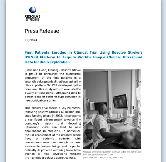
First Patients enrolled in clinical trial using Resolve Stroke’s SYLVER Platform
Resolve Stroke has enrolled the first patients in a groundbreaking clinical trial using its #SYLVER platform to evaluate transcranial ultrasound data for detecting cerebral hypoperfusion in neurocritical care units. This significant milestone brings us closer to revolutionizing the application of ultrasound in neurology, much like the transformative journey of cardiac ultrasound years ago.